Molecular imaging holds immense scientific advantages in preclinical research, revolutionizing the understanding of biological processes at the molecular level. By employing various imaging techniques, such as positron emission tomography (PET), single-photon emission computed tomography (SPECT), and optical imaging, researchers can non-invasively visualize and monitor the molecular interactions, cellular processes, and disease progression within living organisms.This enables a comprehensive assessment of drug pharmacokinetics, therapeutic efficacy, and the evaluation of novel biomarkers. Moreover, the ability to track molecular events longitudinally and dynamically enhances the accuracy and efficiency of preclinical studies, accelerating the development of new treatments and personalized medicine approaches. Through its non-destructive and high-resolution capabilities, molecular imaging empowers researchers to explore the intricate world of molecular biology, unraveling the mysteries of life and advancing scientific knowledge.
"An image is a bridge between the unknown and the known." - Wim Wenders
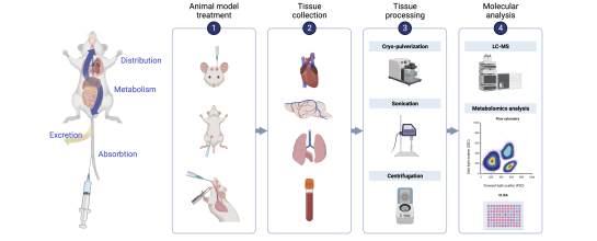
The biodistribution of a new drug is evaluated to understand how the drug is distributed within the body after administration. This information is crucial for assessing its efficacy, safety, and potential toxicities. Several methods can be employed to evaluate the biodistribution and pharmacokinetic profile, of which blood and tissue sampling is the most common.
Tissue sampling involves the collection of various tissues or organs from animals at specific time points after drug administration. These samples can be analyzed using techniques such as high-performance liquid chromatography (HPLC), LC-mass spectrometry (LC-MS), immunohistochemistry, or enzyme-linked immunosorbent assay (ELISA) to determine drug concentrations and localization.
However, this approach is very labor-intensive, expensive, and does not allow for longitudinal studies within the same animal.

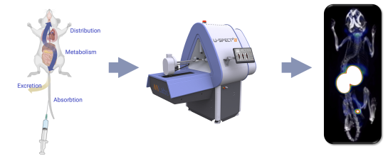
Molecular imaging techniques, such as SPECT, PET, or fluorescence imaging, can be employed to visualize the distribution of drugs, cells or molecules within the body. This approach utilizes the incorporation of imaging agents or fluorescent labels into the compound of interest, allowing for non-invasive imaging and localization within the same animal over time.
Molecular imaging provides faster and better insights in drug targeting and pharmacokinetics, while reducing and refining animal use (3R-principle) in a cost- and time-effective manner.

